See This Report on Spectrophotometers
See This Report on Spectrophotometers
Blog Article
The Ultimate Guide To Spectrophotometers
Table of ContentsSpectrophotometers for BeginnersThe 20-Second Trick For Circularly Polarized LuminescenceHow Spectrophotometers can Save You Time, Stress, and Money.The 30-Second Trick For Uv/visThe 8-Minute Rule for Circular DichroismNot known Facts About SpectrophotometersThe Of Uv/vis6 Simple Techniques For Circular DichroismGetting The Circular Dichroism To WorkUv/vis Fundamentals ExplainedThe Definitive Guide for SpectrophotometersThe 15-Second Trick For Uv/visThe Facts About Uv/vis Uncovered
It is then scanned through the sample and the recommendation options. Fractions of the event wavelengths are transmitted through, or reflected from, the sample and the referral. Electronic circuits convert the relative currents into direct transmission portions and/or absorbance/concentration values.The transmission of a reference compound is set as a baseline (datum) value, so the transmission of all other compounds are tape-recorded relative to the preliminary "zeroed" substance. The spectrophotometer then transforms the transmission ratio into 'absorbency', the concentration of particular components of the test sample relative to the initial compound.
Given that samples in these applications are not easily available in big amounts, they are specifically suited to being analyzed in this non-destructive strategy. In addition, valuable sample can be saved by making use of a micro-volume platform where as low as 1u, L of sample is required for complete analyses. A short explanation of the treatment of spectrophotometry includes comparing the absorbency of a blank sample that does not consist of a colored substance to a sample which contains a colored substance.
The Basic Principles Of Circularly Polarized Luminescence
In biochemical experiments, a chemical and/or physical home is selected and the procedure that is used is particular to that home in order to obtain more info about the sample, such as the amount, purity, enzyme activity, and so on. Spectrophotometry can be used for a variety of methods such as identifying optimum wavelength absorbance of samples, identifying optimum p, H for absorbance of samples, determining concentrations of unknown samples, and identifying the p, Ka of numerous samples.: 21119 Spectrophotometry is also a helpful procedure for protein filtration and can also be utilized as a technique to produce optical assays of a substance.
It is possible to understand the concentrations of a 2 part mix utilizing the absorption spectra of the standard services of each part. To do this, it is needed to know the extinction coefficient of this mixture at two wave lengths and the extinction coefficients of services that include the known weights of the 2 parts.
Some Known Facts About Uv/vis.
Many spectrophotometers are used in the UV and noticeable regions of the spectrum, and a few of these instruments likewise run into the near-infrared region too. The concentration of a protein can be approximated by measuring the OD at 280 nm due to the existence of tryptophan, tyrosine and phenylalanine (https://pxhere.com/en/photographer/4182440).
This approach requires a spectrophotometer capable of determining in the UV area with quartz cuvettes.: 135 Ultraviolet-visible (UV-vis) spectroscopy involves energy levels that delight electronic transitions. Absorption of UV-vis light excites particles that are in ground-states to their excited-states.
20. 8 O.D. Ink makers, printing companies, fabrics vendors, and much more, require the information supplied through colorimetry. They take readings in the region of every 520 nanometers along the visible area, and produce a spectral reflectance curve or a data stream for alternative discussions. These curves can be used to check a new batch of colorant to check if it makes a match to specs, e.
Facts About Circularly Polarized Luminescence Revealed
Conventional visible area spectrophotometers can not detect if a colorant or the base product has fluorescence. This can make it difficult to handle color issues if for instance several of the printing inks is fluorescent. Where a colorant includes fluorescence, a bi-spectral fluorescent spectrophotometer is used (https://www.kickstarter.com/profile/olisclarity1/about). There are two significant setups for visual spectrum spectrophotometers, d/8 (round) and 0/45.
Researchers utilize this instrument to determine the quantity of compounds in a sample. If the substance is more concentrated more light will be soaked up by the sample; within small varieties, the Beer, Lambert law holds and the absorbance in between samples differ with concentration linearly. In the case of printing measurements two alternative settings are frequently used- without/with uv filter to control better the impact of uv brighteners within the paper stock.
Getting My Circular Dichroism To Work
Some applications require little volume measurements which can be performed with micro-volume platforms. As explained in the applications section, spectrophotometry can be used in both qualitative and quantitative analysis of DNA, RNA, and proteins. Qualitative analysis can be utilized and spectrophotometers are utilized to tape spectra of compounds by scanning broad wavelength areas to figure out the absorbance residential or commercial properties (the strength of the color) of the substance at each wavelength.
Indicators on Circularly Polarized Luminescence You Should Know
One major element is the kind of photosensors that are offered for different spectral areas, but infrared measurement is also tough due to the fact that practically everything releases IR as thermal radiation, specifically at wavelengths beyond about 5 m. Another issue is that many products such as glass and plastic soak up infrared, making it incompatible as an optical medium.
Samples for IR spectrophotometry may be smeared between two discs of potassium bromide or ground with potassium bromide and pushed into a pellet. Where liquid options are to be measured, insoluble silver chloride is used to build the cell. Spectroradiometers, which operate practically like the noticeable area spectrophotometers, are developed to determine the spectral density of illuminants. Recovered Dec 23, 2018. Basic Laboratory Methods for Biochemistry and Biotechnology (Second ed.). The necessary guide to analytical chemistry.
Oke, J. B.; Gunn, J. E.
Spectrophotometers - Truths
Ninfa AJ, Ballou DP, Benore M (2015 ). Fundamental Laboratory Techniques for Biochemistry and Biotechnology (3, rev. ed.). UV/Vis. Laboratory Devices.
Everything about Spectrophotometers
Recovered Jul 4, 2018. Trumbo, Toni A.; Schultz, Emeric; Borland, Michael G.; Pugh, Michael Eugene (April 27, 2013). "Applied Spectrophotometry: Analysis of a Biochemical Mixture". Biochemistry and Molecular Biology Education. 41 (4 ): 24250. doi:10. 1002/bmb. 20694. PMID 23625877. (PDF). www. mt.com. Mettler-Toledo AG, Analytical. 2016. Retrieved Dec 23, 2018. Cortez, C.; Szepaniuk, A.; Gomes da Silva, L.
"Exploring Proteins Filtration Strategies Animations as Tools for the Biochemistry Mentor". Journal of Biochemistry Education. 8 (2 ): 12. doi:. Garrett RH, Grisham CM (2013 ). Biochemistry. Belmont, CA: Cengage. p. 106. ISBN 978-1133106296. OCLC 801650341. Vacation, Ensor Roslyn (May 27, 1936). "Spectrophotometry of proteins". Biochemical Journal. 30 (10 ): 17951803. doi:10. 1042/bj0301795.
PMID 16746224. Hermannsson, Ptur G.; Vannahme, Christoph; Smith, Cameron L. C.; Srensen, Kristian T.; Kristensen, Anders (2015 ). "Refractive index dispersion picking up utilizing a selection of photonic crystal resonant reflectors". Applied Physics Letters. 107 (6 ): 061101. Bibcode:2015 Ap, Ph, L. 107f1101H. doi:10. 1063/1. 4928548. S2CID 62897708. Mavrodineanu R, Schultz JI, Menis O, eds.
What Does Circularly Polarized Luminescence Do?
U.S. Department of Commerce National Bureau of Standards special publication; 378. Washington, D.C.: U.S. National Bureau of Standards. p. 2. OCLC 920079.
The process starts with a controlled light source that brightens the evaluated sample. In the case of reflection, as this light engages with the sample, some is Learn More Here absorbed or released. The produced light journeys to the detector, which is analyzed, measured, and provided as industry-standard color scales and indices.
Industry governing bodies typically define specific metrics for specific products, such as Tomato and Coffee indices. The streamlined math appears like this: Where R is the reflection coefficient. All terms are examined over the noticeable spectrum from 400 to 700 nm. When it comes to transmission, when the light connects with the sample, it is either taken in, reflected, or sent.
Some Of Circular Dichroism
Examples include APHA (American Public Health Association) for watercolor and purity analysis, ASTM D1500 for petrochemical color analysis, edible oil indices utilized in food, and color analyses of beverages. The simplified mathematics appears like this:. Where T is the transmission coefficient. All terms are examined over the noticeable spectrum from 400 to 700 nm.
Image Credit: Matej Kastelic/ Dr. Arnold J. Beckman and his associates at the National Technologies Laboratories first created the spectrophotometer in 1940. In 1935 Beckman established the business, and the discovery of the spectrophotometer was their most ground-breaking development. Dr. Bruce Merrifield, a Nobel prize-winning biochemist, stated that the development of the spectrophotometer was "probably the most crucial instrument ever developed towards the advancement of bioscience." Before the discovery of the spectrophotometer, chemical analyses took weeks to finish, with 25% accuracy.
Things about Uv/vis
99% accuracy. Gradually, scientists kept improving the spectrophotometer style to enhance its performance. The UV abilities of the model B spectrophotometer were enhanced by changing the glass prism with a quartz prism. Eventually, the Model DU was produced, including a hydrogen lamp and other enhancements. This instrument was used in commercial laboratories, centers, and chemistry and biochemistry departments.
After 1984, double-beam variations of the gadget were designed. The addition of external software with the provision of onscreen display screens of the spectra can be found in the 1990s. Typically, a spectrophotometer is made up of 2 instruments, namely, a spectrometer and a photometer. A fundamental spectrophotometer consists of a light, a monochromator, a collimator for straight light beam transmission, a cuvette to place a sample, and a photoelectric detector.
Indicators on Uv/vis/nir You Need To Know
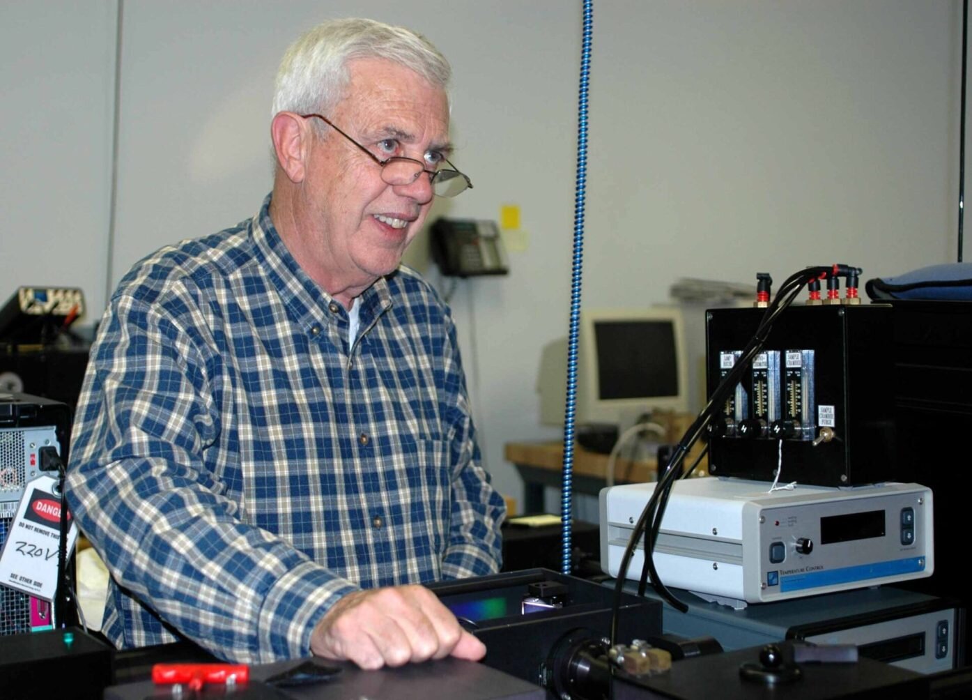
There are various kinds of spectrophotometers in numerous sizes and shapes, each with its own purpose or performance. A spectrophotometer identifies just how much light is shown by chemical parts. spectrophotometers. It measures the distinction in light strength based upon the total amount of light presented to a sample and the quantity of beam that travels through the sample option
Based on the instrument's design, the sample is placed between the spectrometer and the photometer. After the light is travelled through the sample, the photometer determines its strength and displays the reading. A spectrophotometer is used to identify the concentration of both colorless and colored solutes in a solution. This instrument is utilized to figure out the rate of a reaction.
Report this page